Enabling high-resolution, large-scale 3D imaging with LifeCanvas Technologies
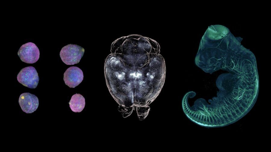
Figure 1: Three types of 3D biological specimens. From organoids to whole organs and embryos, the ability to visualize biological samples in their native three-dimensional architecture has become increasingly important across life science research.
1. Introduction
The ability to visualize biological specimens in three dimensions has become increasingly important across many areas of life science research. From neuroscience and developmental biology to oncology and organoid research, understanding the spatial organization of cells and tissues often requires imaging beyond the limitations of conventional two-dimensional microscopy and two-dimensional biological models (Figure 1).
This need, however, comes with important challenges. Imaging a biological specimen in three dimensions requires not only an appropriate imaging technology, but also a sample that can be effectively accessed by light. Unlike thin, transparent samples, intact tissues and organs are highly heterogeneous and can strongly scatter and absorb light. This limits the penetration of both excitation and emitted fluorescence, making it difficult to obtain high-quality images deep inside the specimen. In addition, labeling large samples uniformly can be challenging, as antibodies and other molecular probes need to reach structures throughout the entire volume.
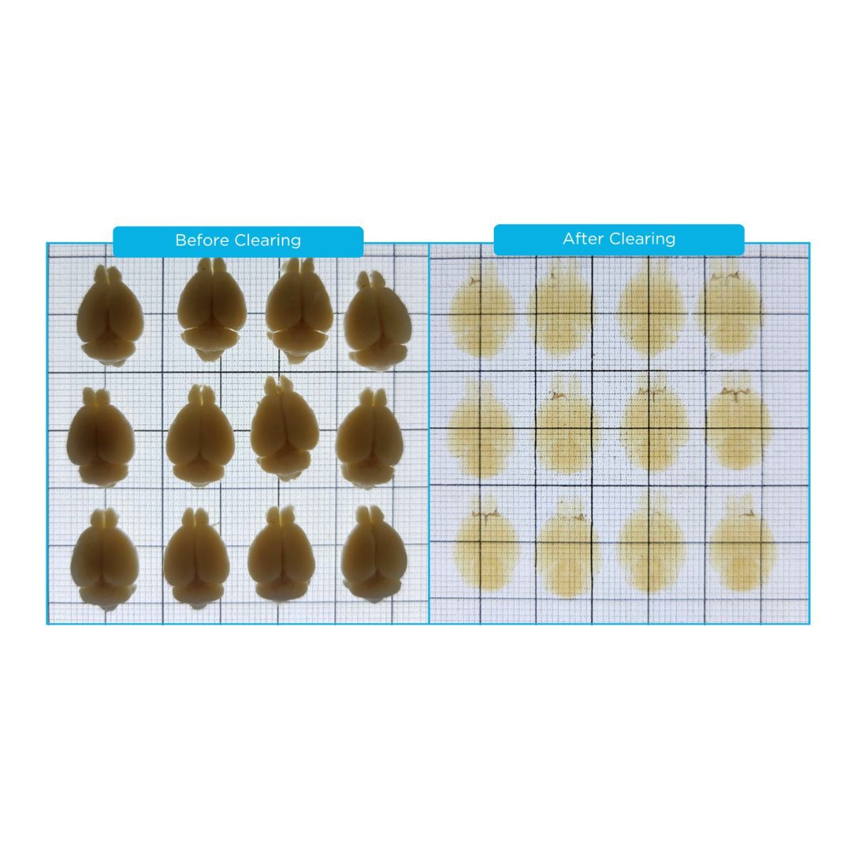
In recent years, the development of tissue-clearing methods has helped overcome many of these limitations by rendering large biological specimens optically transparent. By reducing light scattering and improving optical compatibility between the different components of the sample, tissue clearing has made it possible to image intact organs, whole brains, thick tissue sections and other large biological specimens at cellular resolution. This has opened the way to volumetric imaging approaches capable of preserving and revealing the three-dimensional organization of biological systems.
Once sample transparency could be achieved, the remaining challenge was to develop imaging technologies capable of efficiently capturing these large volumes while maintaining high spatial resolution throughout the specimen. This is where Light Sheet Fluorescence Microscopy (LSFM) has emerged as one of the most powerful approaches for large-scale 3D imaging. Unlike conventional widefield or point-scanning fluorescence microscopy, LSFM illuminates the specimen only within the plane being imaged. A thin sheet of excitation light is introduced into the sample, while fluorescence is collected along an axis orthogonal to the illumination path. This geometry provides efficient optical sectioning while minimizing unnecessary excitation of regions outside the imaging plane.
These features make Light Sheet Microscopy particularly attractive for large-volume imaging. Entire organs, cleared brains, tissue sections, organoids and three-dimensional biological models can be acquired at high speed while limiting photobleaching and phototoxicity. However, not all light sheet microscopes generate and use the illumination sheet in the same way. In this application note, we will explore how LifeCanvas Technologies has addressed these challenges by implementing Axial Sweeping technology to enable high-resolution, large-scale 3D imaging.
2. Axial Sweeping
The main advantage of Light Sheet Microscopy is its selective illumination: a thin sheet of excitation light illuminates only the plane being imaged, while fluorescence is collected perpendicular to the illumination path. This reduces background and unnecessary exposure of the sample, while allowing rapid acquisition of optical sections.
However, generating a thin and uniform light sheet over a large field of view is challenging. In a conventional light sheet, the beam is thinnest at its focal point, where axial resolution is highest. As the beam propagates away from this point, it gradually becomes thicker, leading to a decrease in optical sectioning. As a result, resolution can vary across the field of view. This limitation becomes particularly relevant when imaging large cleared tissues, where a large field of view is needed without compromising image quality.
Axial Sweeping addresses this problem by dynamically moving the narrowest part of the light sheet across the field of view. The movement of the excitation beam is synchronized with image acquisition, so that the camera preferentially collects fluorescence from the region where the light sheet is optimally thin. In LifeCanvas systems, this synchronization is achieved using a rolling-shutter sCMOS camera. As the light sheet sweeps through the sample, the active detection region of the camera follows its position. This allows the system to maintain more consistent optical sectioning across the entire field of view (Figure 2).
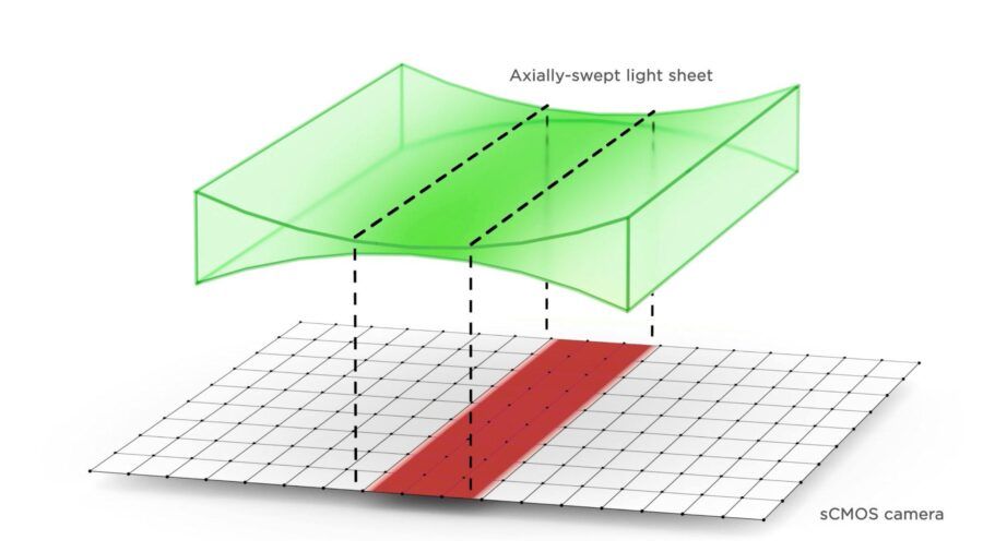
Figure 2: Schematic representation of LifeCanvas’ Axial Sweeping technology, in which the narrowest part of the light sheet is synchronized with the rolling shutter of an sCMOS camera.
In simple terms, while a conventional light sheet has a fixed region of optimal resolution, Axial Sweeping moves this optimal region throughout the sample. The result is a more uniform axial resolution across large fields of view, an important advantage when imaging large biological specimens and performing quantitative 3D analysis (Figure 3).
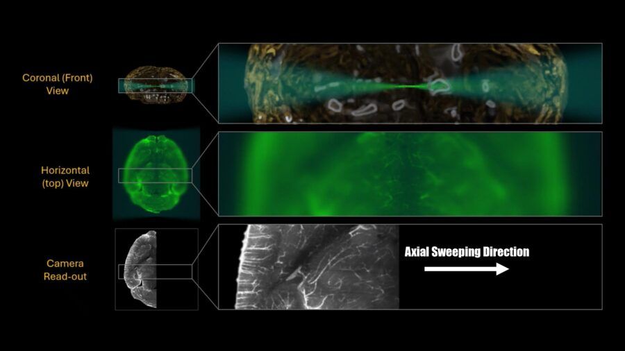
Figure 3: Representation of the effects of Axial Sweeping on image quality. Across the three rows, it can be appreciated that the highest image quality is achieved only at the narrowest part of the light sheet, while the image becomes progressively blurred away from this region. The rolling-shutter camera captures only the optimal region of the light sheet, ensuring that only the highest-quality signal contributes to the final image.
LifeCanvas Technologies has developed its light sheet microscopy portfolio around this concept, combining Axial Sweeping with optical, mechanical, and sample-handling architectures designed for different experimental scales. The result is a portfolio comprising three specific systems: SmartSPIM, MegaSPIM, and DALISPIM.
3. SmartSPIM
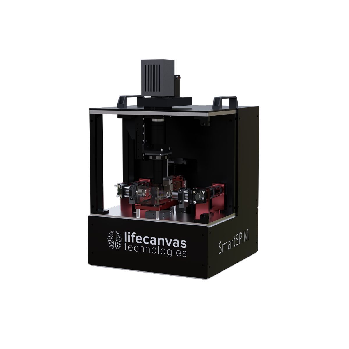
SmartSPIM is designed primarily for high-resolution imaging of large, intact biological specimens. The system is particularly well suited to cleared organs, tissues, and organoids where the goal is to obtain a three-dimensional representation of the specimen while preserving cellular-scale information (Figure 4).
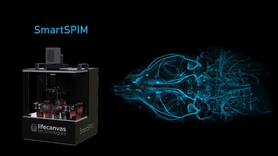
Figure 4: Photograph of the SmartSPIM system and a mouse head image acquired using this light sheet microscope.
At the heart of SmartSPIM is LifeCanvas’ implementation of Axial Sweeping, which enables a homogeneous axial point spread function across the field of view and addresses one of the principal limitations of conventional static light sheet illumination.
SmartSPIM features a flexible imaging chamber capable of accommodating samples ranging from organoids and tissue sections to substantially larger intact specimens. Depending on the configuration, LifeCanvas specifies sample dimensions of up to approximately 40 × 75 mm. The system also supports multiple detection objectives, providing flexibility between large-scale overview imaging and higher-resolution acquisition.
Acquisition speed is another important component of the SmartSPIM design. Large cleared samples can generate extremely large datasets, and acquisition time can quickly become a limiting factor for experimental throughput. SmartSPIM offers acquisition speeds of up to 20 frames per second and, under specified imaging conditions, can acquire a mouse brain hemisphere or similarly sized specimen in less than 30 minutes.
The combination of Axial Sweeping, large sample compatibility, rapid acquisition, and flexible magnification makes SmartSPIM particularly well suited to applications in which intact biological structures need to be interrogated across an extended three-dimensional volume.
Rather than optimizing exclusively for either resolution or sample size, SmartSPIM is designed to provide a practical balance between the two, allowing researchers to move from cleared tissue to a high-resolution volumetric dataset within a single imaging workflow.
4. MegaSPIM
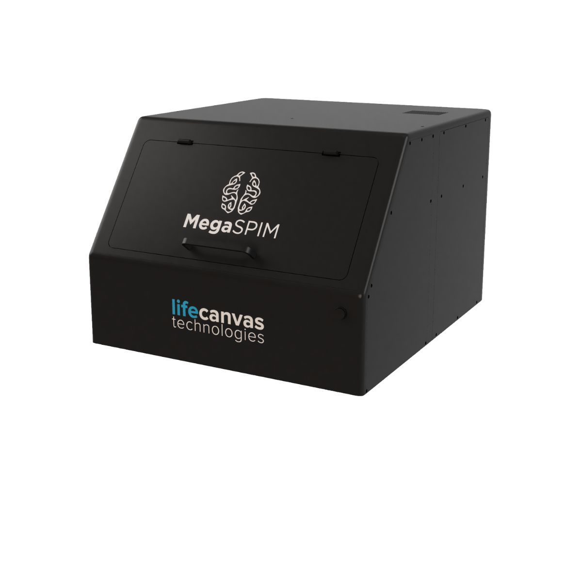
MegaSPIM addresses another major challenge in large-scale light sheet microscopy: imaging extremely large areas in the lateral dimensions. The microscope is designed for samples with lateral dimensions of up to approximately 200 × 200 mm, making it particularly well suited for large tissue sections, tissue slabs, expanded specimens, and arrays containing multiple samples (Figure 5).
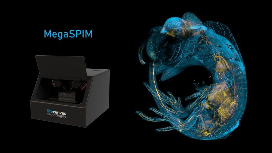
Figure 5: Photograph of the MegaSPIM system and an image of a whole lizard embryo acquired using this light sheet microscope. MegaSPIM is particularly well suited for imaging large biological specimens.
At this scale, field of view becomes a defining parameter of the microscope. Simply increasing the illuminated area, however, does not inherently solve the underlying optical challenge of maintaining a uniform light sheet thickness. MegaSPIM therefore retains Axial Sweeping as a central component of its imaging architecture, allowing the optimal portion of the excitation beam to be dynamically swept across the field rather than relying on a single static beam waist.
The optical architecture of MegaSPIM is specifically adapted to large-format imaging. The system uses long-working-distance objectives positioned at approximately 45° relative to the sample plane. This configuration allows the microscope to cover very large lateral areas while maintaining the geometry required for effective light sheet illumination and fluorescence detection.
MegaSPIM can therefore be considered a platform designed around spatial scale. While SmartSPIM emphasizes high-resolution volumetric imaging of intact samples, MegaSPIM extends the same Axial Sweeping principle to applications in which the lateral dimensions of the specimen represent the primary imaging challenge. By combining a very large field of view with optimized light sheet illumination, MegaSPIM enables high-quality imaging across large sample areas without compromising the principles of high-resolution light sheet microscopy.
5. DALISPIM
The third member of the LifeCanvas portfolio, DALISPIM, takes a different approach to the scalability of light sheet microscopy.
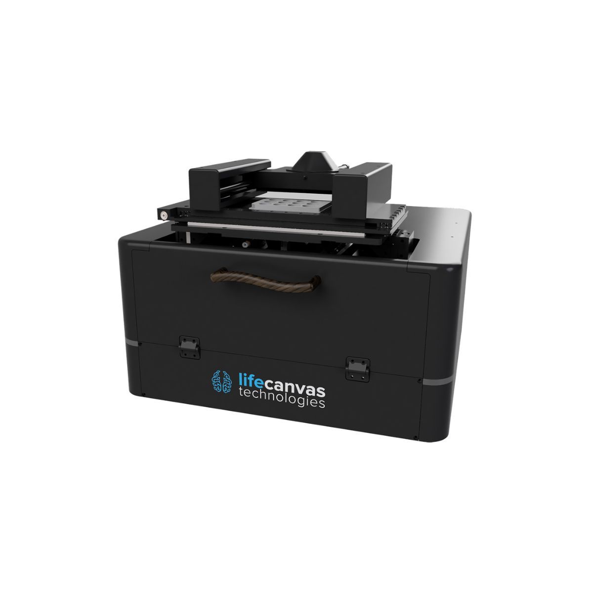
Introduced as an open-top, inverted light sheet microscope, DALISPIM combines high-speed volumetric imaging with flexible sample handling. Its open-top architecture accommodates a broad range of sample formats, including slides, multiwell plates, Petri dishes, suitable for organoids, tissue sections, and other three-dimensional biological models (Figure 6).
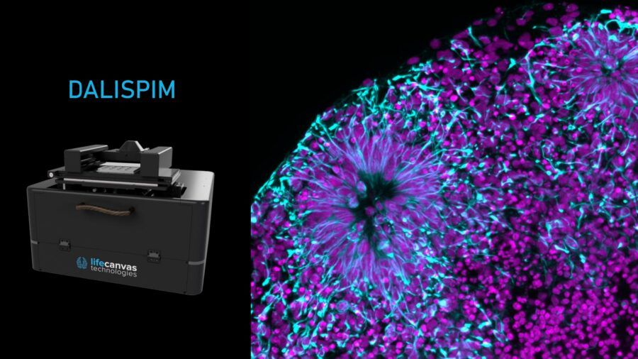
Figure 6: Photograph of the DALISPIM system and an image of an assembloid acquired using this light sheet microscope. This system has been designed for high throughput analysis.
In addition to Axial Sweeping, a key component of DALISPIM is the Depth Adaptive Light Interface (DALI), which maintains an appropriate refractive-index-matched optical interface as imaging progresses through the sample. This is essential for high-resolution imaging, as refractive-index mismatch can introduce aberrations and degrade both illumination and detection.
Together, the open-top configuration and DALI provide the flexibility required for diverse sample formats while maintaining the optical conditions needed for high-quality volumetric imaging. As with the other LifeCanvas systems, Axial Sweeping further enhances optical sectioning by dynamically coordinating illumination and detection across the field of view.
The particular strength of DALISPIM lies in combining this optical performance with a workflow designed for sample diversity and scalability. This makes it particularly well suited to applications such as organoid screens, tissue sections, multiwell experiments, and other high-throughput workflows where large numbers of samples need to be characterized in three dimensions.
DALISPIM therefore complements SmartSPIM and MegaSPIM by addressing a different dimension of scalability: rather than focusing primarily on increasing sample size, it makes the imaging workflow more flexible and scalable.
6. Conclusions
Light Sheet Fluorescence Microscopy has become an essential technology for large-scale three-dimensional imaging because it combines efficient optical sectioning, rapid camera-based acquisition, and reduced excitation outside the imaging plane. These characteristics make LSFM particularly powerful when applied to optically cleared tissues and other large biological specimens.
However, conventional light sheet illumination faces an intrinsic limitation: the excitation sheet cannot remain uniformly thin across an arbitrarily large field of view. As the illuminated region increases, variations in sheet thickness can lead to spatially heterogeneous axial resolution and optical sectioning.
LifeCanvas Technologies’ Axial Sweeping addresses this limitation by transforming the light sheet from a static optical plane into a dynamically scanned imaging region. By synchronizing the movement of the excitation beam with camera detection, the system can continuously position the optimal portion of the light sheet across the field of view. The result is a more homogeneous axial response over a large imaging area, providing a stronger foundation for both visualization and quantitative analysis.
LifeCanvas Technologies has built its light sheet microscopy portfolio around this principle, adapting Axial Sweeping to three complementary imaging platforms. Although these systems address different experimental requirements, they share a common philosophy: large-scale imaging should not require researchers to choose between field of view, resolution, and acquisition speed.
As biological research increasingly moves toward organoids, whole-organ and whole-system analysis, microscopy must evolve accordingly. In this application note, we explored how LifeCanvas Technologies addresses the challenges of imaging large biological specimens through advanced light sheet microscopy solutions. In our next application note, we will focus on sample preparation, showing how LifeCanvas Technologies can support researchers with tissue clearing and labeling workflows to achieve the highest image quality.
Note:
To add more detail to our application note, we want to emphasize that the “Axial sweeping”, was developed by Kevin M. Dean and Reto Fiolka in 2015, please read the articole “Deconvolution-free Subcellular Imaging with Axially Swept Light Sheet Microscopy” to learn more https://www.sciencedirect.com/science/article/pii/S0006349515004981